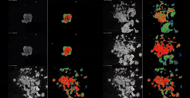
Five threads, one claim: voltage is a control variable for anatomy.
The next 32 slides weave through five themes. Each one earns its place by either grounding the claim in formal language, putting it on trial in a wet lab, or showing what falls out the other side as a tool, a company or a clinic.
01 · Math & morphospace
Form lives in a space.
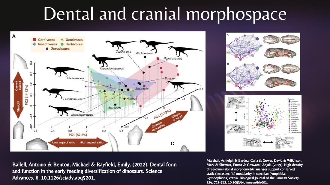
D'Arcy Thompson's geometry, Turing's reaction-diffusion, Raup's three-parameter shell cube, Neural Cellular Automata as Turing's modern descendant, the latent platonic space.
slides 07 · 08 · 09 · 12 · 29
02 · Testable hypotheses
Voltage edits anatomy.
Face-of-a-Frog prepatterns, two-headed planaria, xenobots and anthrobots, oncology — every claim cashed out as a wet-lab perturbation with a readout.
slides 17 · 18 · 19 · 20 · 21 · 22 · 30
03 · Imaging
You can't engineer what you can't measure.
Microelectrodes, vibrating probes, voltage-sensitive dyes, GEVIs, plus FLIM for absolute Vmem in millivolts.
slides 13 · 14 · 15 · 16
04 · Labs
A distributed field, not one lab.
Three featured anchors — Levin / Allen Discovery Center, Wyss Institute, ICDO (Tufts × UVM) — surrounded by Lobo, Bongard / Proteus, Ingber, Cohen, Gentile, Pezzulo. Plus ProbKnow as a live map of the literature and MomBot autonomous wet-lab.
The Bioelectric Code of Life — eight speakers, one day.
Before we get into the foundations: if any of what follows lands, there is a live SEMF workshop
that goes much deeper — bridging fundamental theory with real applications of bioelectric
control and morphogenesis. bioelectricitycourse.com
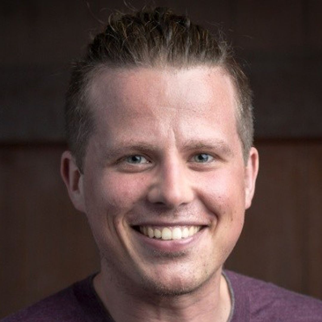
Joel Dietz
MIT · CIMC
Intellectual history · mathematical biology · morphospace theory
Michael Levin
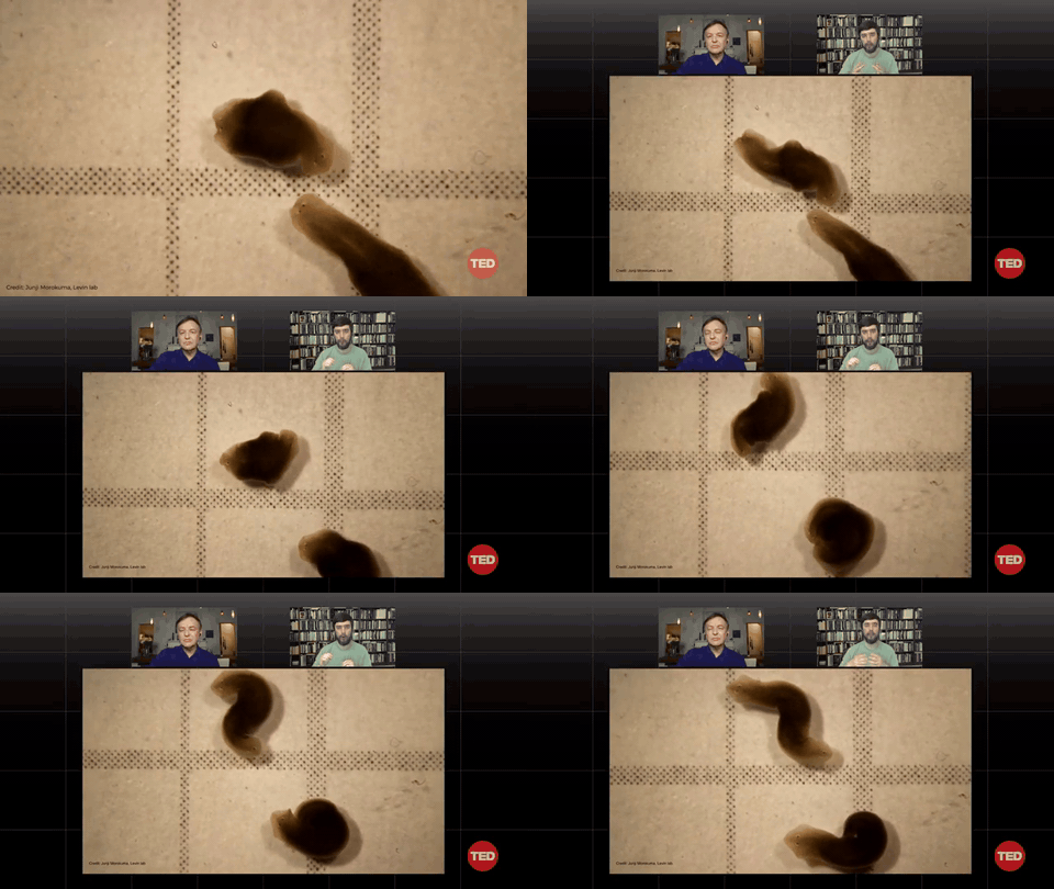
Tufts · Harvard Wyss
Developmental biology · bioelectricity · regenerative medicine
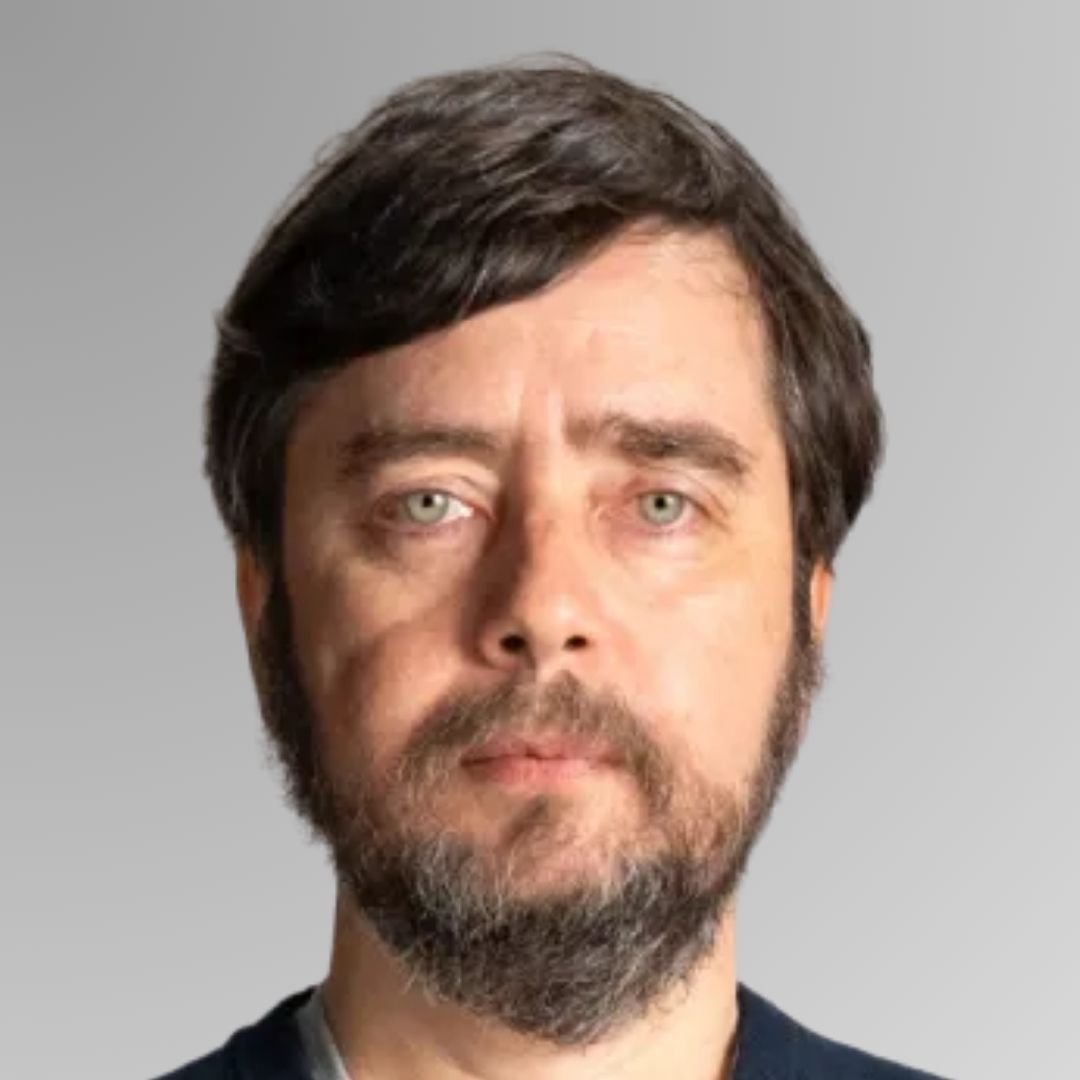
Emmett Shear
Softmax
AI alignment · complex systems · technology entrepreneurship
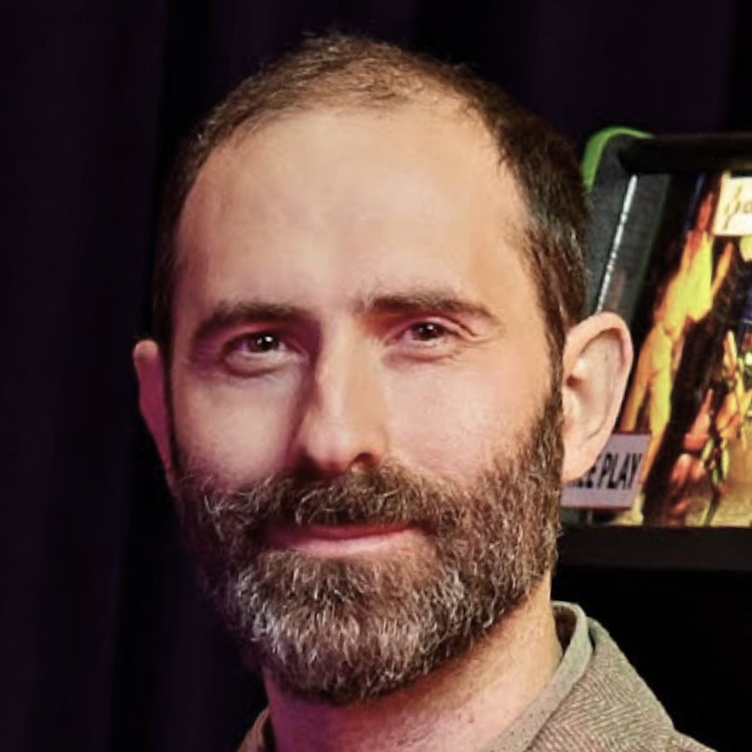
Derek Lomas
TU Delft · Playpower
Positive AI · education · cognitive science
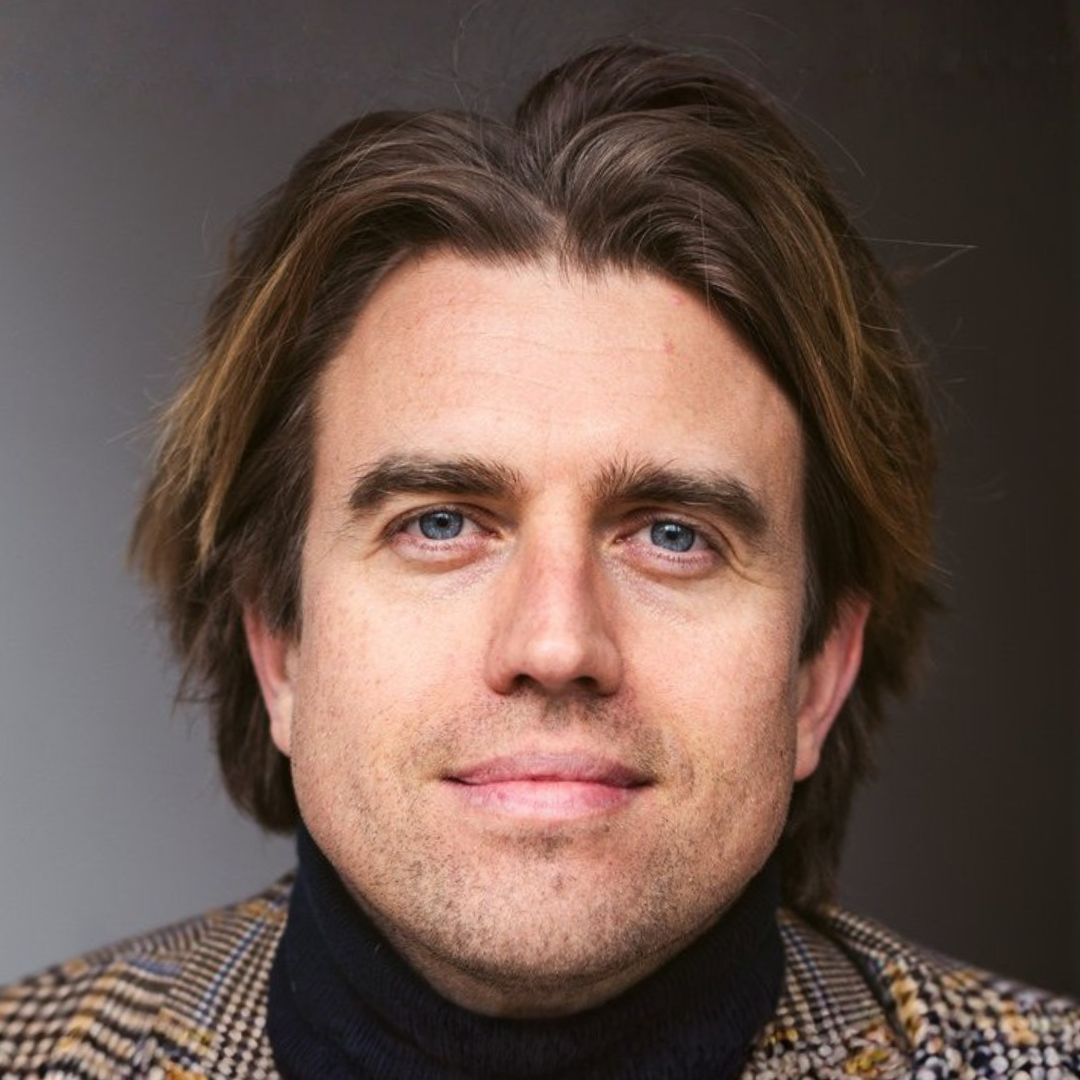
Adam Goldstein
Hum Labs
Multi-agent RL · AI alignment · collective intelligence
Embodied cognition · neuroscience of learning · collective intelligence
03 / 33
Slide 04 · Mapping the literature
Tool · ProbKnow · probknow.com
The field has grown faster than any one researcher can track.
ProbKnow is a probabilistic knowledge graph for bioelectricity research — papers,
researchers, organizations and concepts, all linked by inferred relationships.
Surfaces hidden connections between labs, papers and ideas the citation graph misses.
Lets a non-expert ask "who works on planarian regeneration AND uses GEVIs?" and get a navigable answer.
Updates as new papers land, so the map stays current as the field accelerates.
ProbKnow knowledge graph · live exploration of the bioelectricity literature.
04 / 33
Slide 05 · Key terms in Michael Levin
Glossary · hover any card for the definition
Ten words that do most of the heavy lifting.
Basal Cognition
Levin · Lyon · 2020
Minimal goal-directed problem-solving in non-neural cells, tissues, and even single-celled organisms — intelligence without brains.
hover
Morphogenesis
D'Arcy Thompson · 1917
The process by which a developing organism builds its three-dimensional body shape from a single fertilized cell.
hover
Anatomical Homeostasis
Levin lab · 2010s
Active maintenance and restoration of the correct body structure — the regulator that knows when to stop regenerating.
hover
Multi-Scale Competency Architecture (MCA)
Levin · 2022
Nested problem-solvers — molecules, cells, tissues, organs — each with their own goals; intelligence emerges from agents-of-agents.
hover
Xenobots
Kriegman · Levin · 2020
Programmable living machines built from frog (Xenopus) skin cells — body shape designed in silico, assembled from biological parts.
hover
Anthrobots
Gumuskaya · Levin · 2024
Self-assembling motile spheroids built from human tracheal cells — same idea as xenobots, human substrate, no genetic modification.
hover
Anatomical Compiler
Levin · 2014 →
An aspirational interface: specify a target body plan in software, and a stack translates it into the cellular signals that build it.
hover
Cognitive Light Cone
Levin · 2019
The spatiotemporal scope of an agent's goals — how far in space and time it can represent and act upon outcomes.
hover
Morphospace
Raup · 1966
The abstract space of all possible body plans — most points are empty; evolution and bioelectricity navigate it.
hover
Platonic Spaces
Levin · Hazan · 2025
A latent space of pre-existing patterns biology appears to "download" rather than design — rhymes with morphospace, deeper claim.
hover
05 / 33
Slide 06 · How we discovered cells run on electricity
1780s–1952 · Galvani → Hodgkin & Huxley
It took ~170 years to be sure cells generate their own voltage.
Five experiments settled the basic question — do cells produce electricity at all? Hover any card for the detail.
1780–91
Galvani
hover
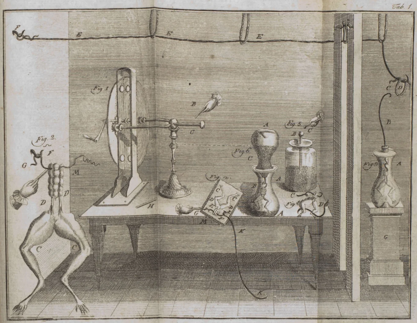
Luigi Galvani — frog leg twitches when touched by a bimetal arc. Calls it animal electricity and proposes the muscle itself is the source.
1800
Volta
hover
Alessandro Volta — argues the current comes from the dissimilar metals, not the frog. Builds the voltaic pile (battery) to prove it.
1840s–50s
du Bois-Reymond
hover
Emil du Bois-Reymond — sensitive galvanometers detect electrical signals in nerve and muscle, including the "negative variation" later understood as the action potential. Galvani vindicated.
1902
Bernstein
hover
Julius Bernstein — membrane theory: cells are polarised because the membrane is selectively permeable to K⁺. Resting potential gets a mechanism.
1939–52
Hodgkin & Huxley
Nobel 1963 · hover
Hodgkin & Huxley — squid giant axon, voltage clamp, four ODEs that quantitatively predict the action potential. The mechanism becomes maths. Nobel 1963
Plate from Galvani's De Viribus Electricitatis (1791) · animated reconstruction of the bimetal-arc circuit closing.
06 / 33
Slide 07 · The thesis, 100 years early
1917
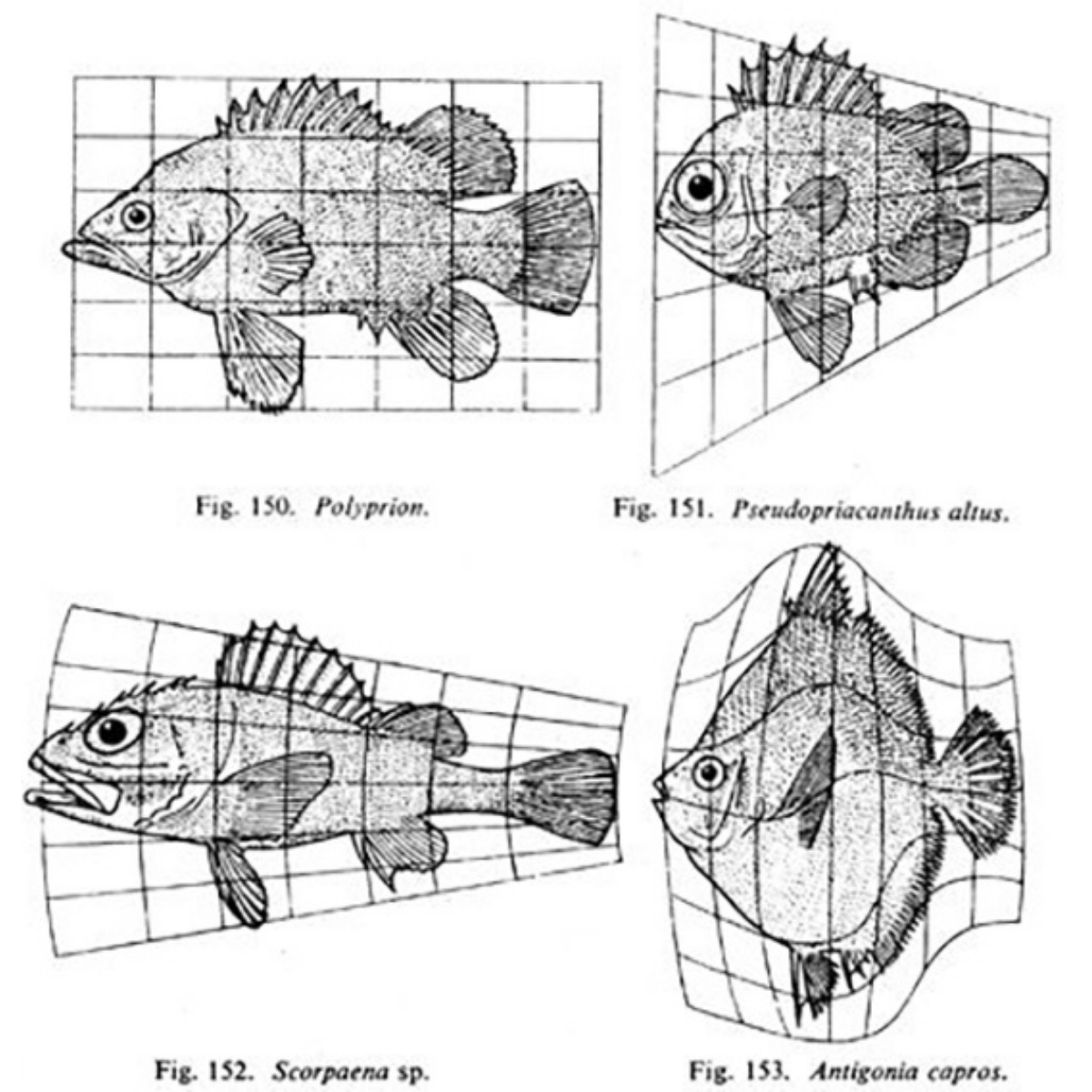
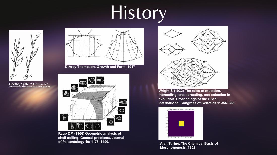
D'Arcy Thompson: form is geometry, not just inheritance.
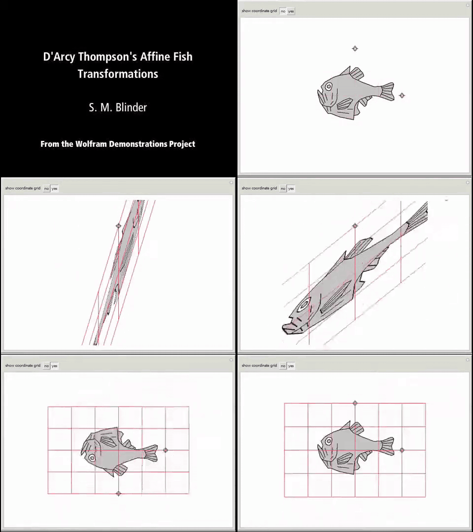
In On Growth and Form, Thompson showed you could turn one species' body into another's by
warping a coordinate grid — not by changing the parts list.
That image is a conceptual precursor to bioelectricity research a century later: there is some
field, some tissue-scale instruction, that helps decide shape on top of the cellular
hardware.
The genome lists ingredients. Something else does the warping.
Original · On Growth and Form, 1917 · Figs. 150–153
Same idea, animated · Wolfram Demonstrations
07 / 33
Slide 08 · Turing's reaction-diffusion
1952 · Alan Turing · The Chemical Basis of Morphogenesis
The math arrived 25 years before the biology.
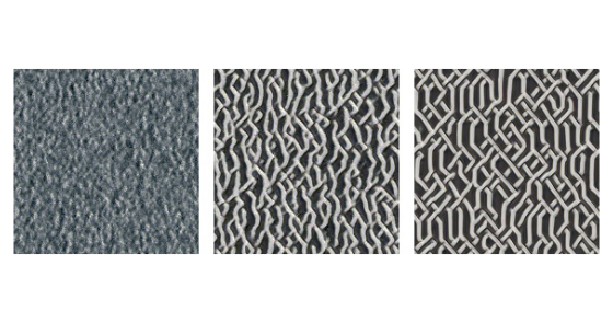
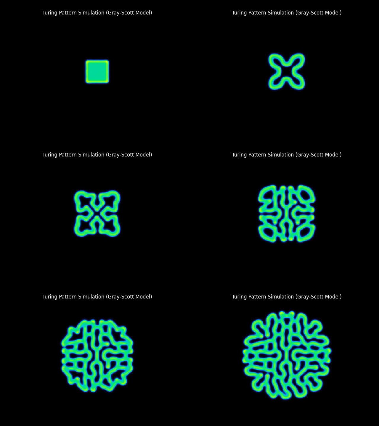
In 1952, less than two years before his death, Turing showed that two diffusing chemicals with the right
nonlinear interaction can spontaneously break a uniform tissue into stable spatial
patterns — spots, stripes, waves. From nothing but the math.
Activator + inhibitor diffusing at different rates — the activator self-amplifies locally, the inhibitor diffuses faster and suppresses neighbours. A uniform field becomes a regular pattern.
Predicted patterns ~25–40 years before any biological mechanism was confirmed.
Vindicated in marine angelfish stripes (Kondo & Asai, Nature 1995), mouse digit spacing (Sheth et al., Science 2012), hair-follicle spacing (Sick et al., Science 2006), and fish skin pigmentation.
Gray-Scott reaction-diffusion simulation (activator V × inhibitor U, du=0.2 / dv=0.1, f=0.055 / k=0.062) — a noisy central seed self-organizes into Turing spots. Rendered from fractastical/morpholib · 1952-turing-morpho.py.
08 / 33
Slide 09 · The space of all possible bodies
1966 · David Raup
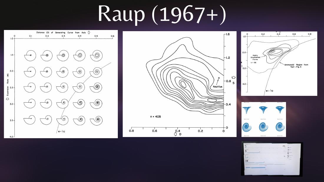
If form is geometry, then there is a space of all forms — and most of it is empty.
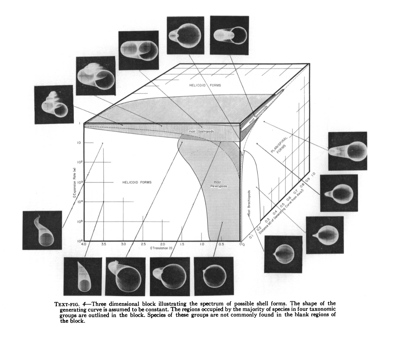
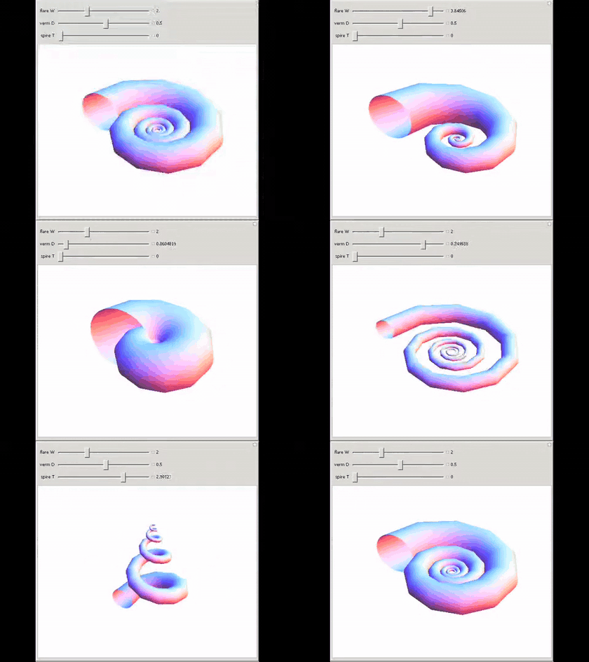
Raup parameterised gastropod shells with just three numbers — W (whorl expansion rate),
D (distance from the coiling axis), and T (translation rate along the axis). Sweep the
parameters and you generate the full theoretical morphospace of coiled shells.
The striking finding: sampled gastropod shells occupy only a small region of that space. Most of the
cube is geometrically possible but biologically unused.
Form lives in a space. Evolution and development pick a trajectory through it.
Raup 1966 · the original W × D × T cube
Raup 1967 · n=405 shell occupancy, W×D plane
Same parameters, animated · Wolfram
09 / 33
Slide 10 · How cells actually get into shape
Primer · the morphogenesis toolkit
Cells build bodies with a small set of repeatable tricks.
Before bioelectricity enters the story, here are the four classical mechanisms developmental
biology uses to explain how a sheet of cells becomes an organ. Bioelectricity sits on top of all of them.
Cells sort themselves by adhesion (Steinberg, 1963 / Glazier-Graner CPM, 1992); voltage prepatterns sit on top of the result.
1
Chemical gradients (morphogens)
Diffusing signal molecules — Wnt, BMP, Sonic hedgehog, retinoic acid — form concentration gradients across tissue. Cells read their position in the gradient and switch on different genes (Wolpert's positional information / French-flag model, 1969).
2
Differential adhesion
Cells expressing different cadherins stick more strongly to "like" cells. Mixed populations spontaneously sort into nested layers — Steinberg's differential adhesion hypothesis (1963). Drives gastrulation and tissue boundaries.
3
Mechanical forces & cell shape
Actomyosin contraction at the apical surface (apical constriction) folds sheets into tubes; convergent extension elongates the embryo. The neural tube, gut and heart all form by coordinated mechanics, not just signalling.
4
Bioelectric prepatterns & stigmergy
Tissue-scale voltage maps (Vmem), shared via gap junctions, can bias migration, organ shape and gene expression — sometimes upstream of the chemical and mechanical layers. A bioelectric prepattern is one of these maps laid down before cells visibly differentiate — a stigmergic medium for shape. The next slides go deep on this one.
10 / 33
Slide 11 · The mechanism in 90 seconds
Primer · how bioelectricity actually works
Cells run on voltage — and they share it across tissue.
Three ingredients turn single-cell voltages into a tissue-scale instruction. Every result on the next five slides is a read or a write on this layer.
1
Every cell has a voltage (Vmem)
Ion pumps and channels move K⁺, Na⁺ and Cl⁻ across the membrane. Net result: tens of millivolts (Vmem) across the membrane of every cell — not just neurons.
2
Cells share voltage via gap junctions
Gap junctions are direct ion channels between neighbouring cells. Patches settle into shared voltage states, building a tissue-scale bioelectric pattern over hours.
3
The pattern instructs anatomy
Vmem patterns can gate downstream gene expression. In several systems, induced patterns bias anatomical outcomes. Read with voltage dyes; write with channel openers/blockers.
11 / 33
Slide 12 · The computational mirror
2020–2021 · Mordvintsev, Randazzo, Niklasson, Levin · Distill · Google Research
The same answer, in silicon: local rules → global form.i
what's an NCA?
If Turing showed that local diffusion rules can spontaneously produce stable patterns, a recent line of work shows the same thing with learned local rules. Neural Cellular Automata are tiny neural networks where every "cell" runs the same update rule on its own state plus its neighbours. Train them, and they grow lizards from a single seed pixel, regenerate after damage, classify MNIST digits collectively, or self-organise complex textures — all from purely local interactions.
Growing NCA · Mordvintsev, Randazzo, Niklasson & Levin, Distill 2020 — live embed. Scroll inside the frame to see lizards grown from a single seed pixel and regenerated after damage.
Bioelectric Vmem patterns are doing the analog version of what NCAs do in silico — every cell runs the same local rule on its own membrane voltage + its neighbours' (gap-junction-coupled). Substrate differs (ion channels vs learned weights); the math is the same. Levin co-authors the Distill series.
12 / 33
Slide 13 · How we actually see voltage in tissue
Primer · the imaging toolkit
Four generations of probes for reading Vmem — and one calibration trick.
Every result on the next slides depends on a measurement technique. The field grew when the tools moved from
one cell at a time to a glowing map of a whole embryo.
1
Microelectrodes & patch clamp
Hodgkin & Huxley, 1949–52 (squid axon) → Neher & Sakmann, 1976 (patch clamp, Nobel 1991). Sharp microelectrodes impale single cells to read Vmem in millivolts; patch clamp seals onto a membrane patch to resolve single ion channels. Gold standard for accuracy — but one cell at a time, and invasive.
2
Vibrating probe (extracellular)
Jaffe & Nuccitelli, 1974 (J. Cell Biol.). A platinum ball oscillates near tissue, mapping endogenous ion currents in the bath without touching cells. Revealed steady currents in eggs, regenerating limbs and wounds — first quantitative window onto developmental bioelectricity.
3
Voltage-sensitive dyes (VSDs)
Cohen, Salzberg & Davila, 1973 — fluorescent small molecules whose brightness tracks Vmem. Adapted for embryos by Vandenberg, Morrie & Adams, 2011 (Dev. Dyn.) and protocolised by Adams & Levin, 2012 using CC2-DMPE + DiBAC4(3) as complementary dyes (one cationic, one anionic — read jointly, not as a designed FRET pair). This is what produced the "Face of a Frog" map on the next slide.
4
Genetically encoded voltage indicators
ArcLight (Jin et al., 2012) → ASAP family (St-Pierre et al., 2014) → Archon (Piatkevich et al., 2018) → Voltron (Abdelfattah et al., 2019). Fluorescent-protein sensors expressed in chosen cells — voltage readout becomes cell-type specific and works in vivo, in flies, fish and mice.
13 / 33
Slide 14 · A history of bioelectric imaging
1791 → today · the instruments that made the field
Each new instrument opened a new biology.
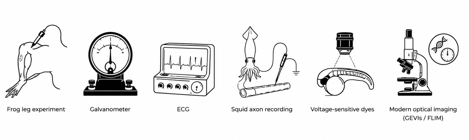
Bioelectricity has always been a measurement-limited field — from the first detection of an animal current to whole-embryo voltage movies.
Frog leg → galvanometer → ECG → squid axon → voltage-sensitive dyes → modern optical imaging (GEVIs / FLIM)
1791
Frog leg experiment · Galvani · twitches establish animal electricity. The field exists.
1840s
Galvanometer · Matteucci, Du Bois-Reymond · sensitive needle/coil instruments make biological currents quantitative for the first time.
1887 / 1903
ECG · Waller, Einthoven · the string galvanometer makes the heart's electrical signal a routine clinical readout. (Nobel 1924.)
1939–52
Squid axon recording · Hodgkin & Huxley · intracellular recording → voltage clamp → quantitative ionic-current model of the action potential. Excitable cells become a math problem. (Nobel 1963.)
1972–77
Voltage-sensitive dyes · Cohen, Salzberg, Davila et al. · merocyanine 540 and successors let you see voltage as light — first in axons, then in many cells at once. Voltage becomes an image.
1997 → today
Modern optical imaging · GEVIs & FLIM · genetically encoded voltage indicators (FlaSh 1997 → ASAP / Archon / Voltron) target specific cell types in vivo; FLIM readouts (2015–19) deliver absolute Vmem in millivolts. This is the instrument set that makes Levin-style developmental bioelectricity quantitative.
From a relative thermometer to an absolute Vmem ruler.
Brightness drifts with loading, expression, bleaching and laser power. Lifetime — the nanoseconds the dye spends in the excited state — doesn't. Read the lifetime of the same dye/GEVI and you get voltage in actual millivolts: "this cell is at −42 mV."
Relative — drifts with loading, bleaching, laser power. Not in mV.
Lifetime · nanoseconds
−42 mV
Absolute — independent of loading, bleaching, laser. Voltage in millivolts.
15 / 33
Slide 16 · Plasticity & learning in cultured networks
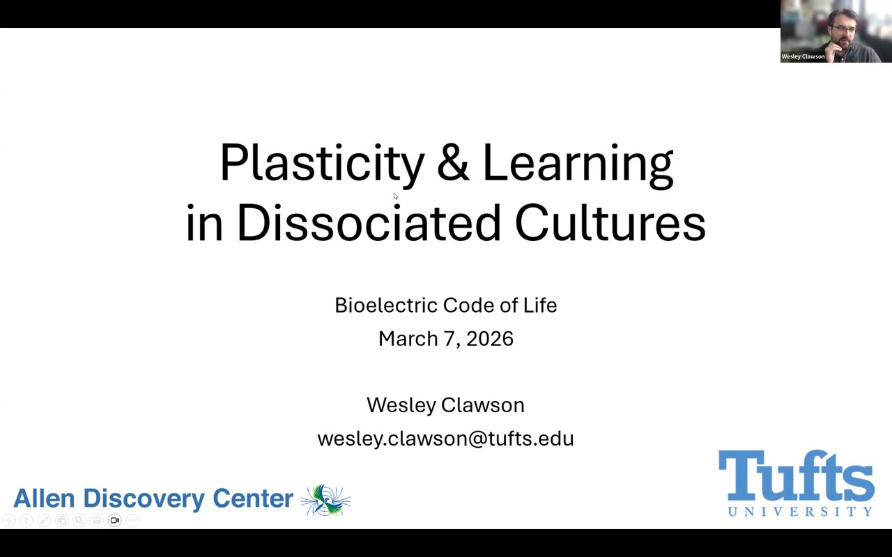
Wesley Clawson · Levin lab / Allen Discovery Center · 2024 → present
Are dissociated neural cultures trainable at the network level?
Wesley Clawson brings closed-loop electrophysiology into dissociated cultures to ask whether learning rules — hebbian, homeostatic, burst-dependent STDP, network-level attractor reshaping, credit assignment — emerge in tissue stripped of body and brain. Two decades of work say cultures are highly adaptable, possibly controllable collectives. The open question is which rule dominates.
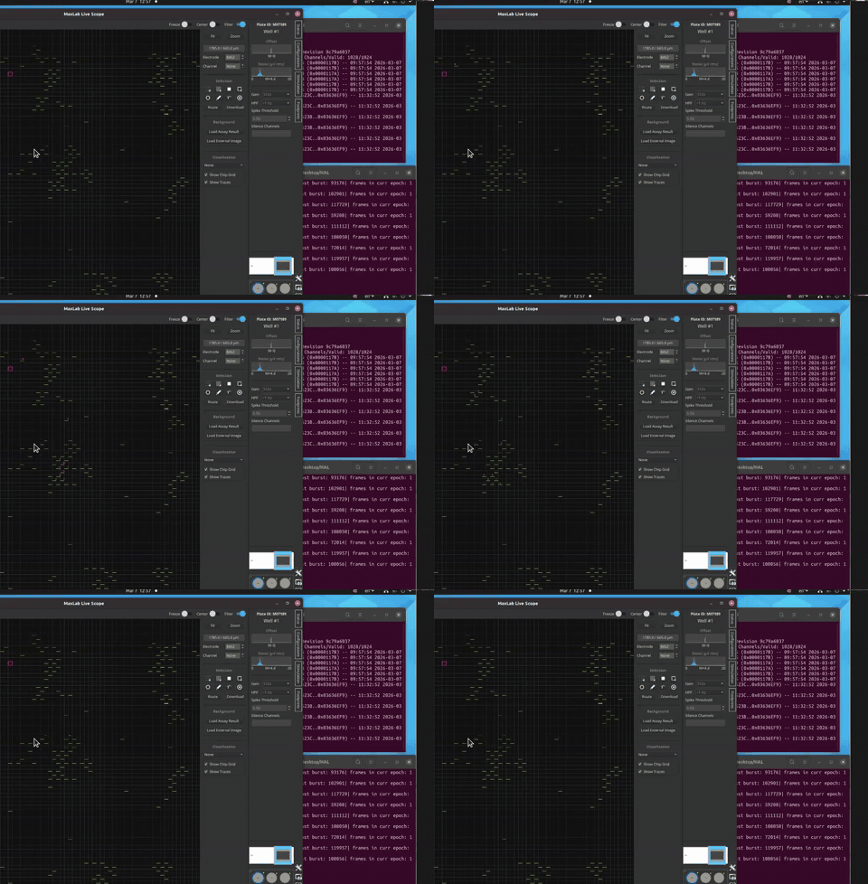
MaxLab Live Scope · CMOS-MEA grid (left) shows spatial spike events; right panel scrolls per-burst frame counts as the network fires.
The setup — dissociated cultures grown on multi-electrode arrays, driven through closed-loop hardware (cf. DishBrain · Cortical Labs · FinalSpark on slide 23). Wesley's twist: ask whether the bioelectric repertoire itself changes under stimulation — not just spike patterns, but the rules.
16 / 33
Slide 17 · First direct evidence
2011 · Vandenberg, Morrie & Adams · Levin lab
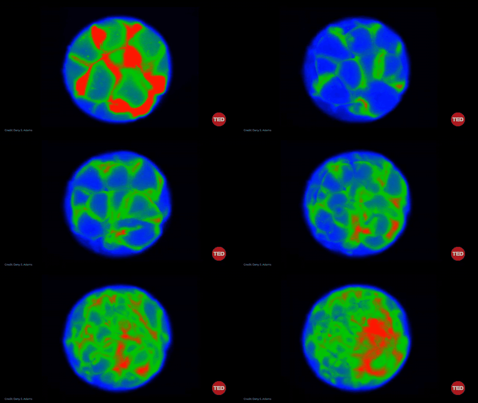
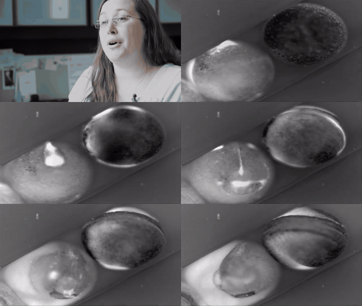
The face appears as a voltage map before any features form.
Using voltage-sensitive dye, the Levin lab imaged the surface of Xenopus
embryos as the head developed. Hyper- and depolarized regions pre-figured the eyes, mouth,
and nostrils — sometimes hours before the cells got there.
Disrupt the voltage pre-pattern and you get craniofacial mispatterning (colloquially the
"Picasso tadpole" phenotype) — features in the wrong places.
Bioelectric prepatterns predict and influence the craniofacial pattern that follows.
Voltage map · CC2-DMPE + DiBAC4(3)
Anatomy follows · brightfield time-lapse
Modern follow-up · 2026 · post-wound wave-vector flow
17 / 33
Slide 18 · The blueprint is rewritable
2010–17 · Oviedo, Beane, Durant, Levin et al.
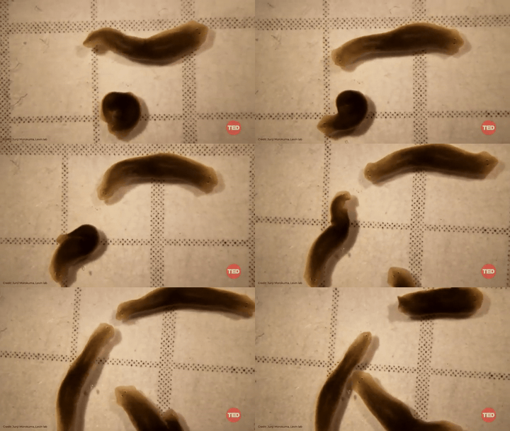
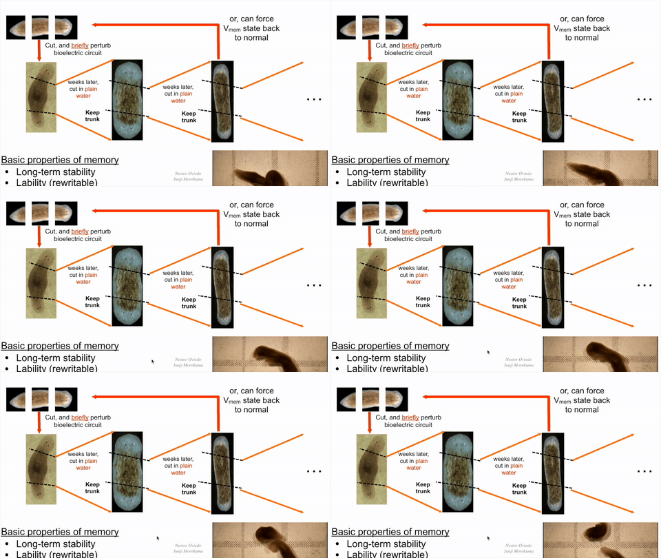
Two heads, same DNA — and the change persists.
Cut a planarian flatworm in half and it normally regenerates one head and one tail. Briefly
perturb its bioelectric circuit before cutting and it can grow two heads instead — and
in Durant et al. (2017) that the altered head/tail target morphology is maintained across subsequent re-amputations via stable gap-junctional state changes,
even though the genome was never touched.
Body shape is partly stored in tissue-level voltage gradients (Vmem)
That store is rewritable and the rewrite can persist across regeneration
It behaves like pattern memory: long-term, addressable, content-specific
Live regeneration footage
The bioelectric edit · two-headed phenotype
18 / 33
Slide 19 · Bodies as graphs, regeneration as rewrites
Lobo · Malone · Levin · 2013 → 2015
If a body plan is a graph, regeneration is a graph-rewriting rule.
1 · Body → Graph
Anatomy collapses to a labelled directed graph: regions are nodes, adjacency is edges.
2 · Cut = graph rewrite
An anterior cut is a rewriting rule: split the graph; each fragment must regenerate its missing nodes.
3 · Regrowth = run the grammar
Lobo & Levin 2015 evolved a regulatory network whose grammar reproduces every documented planarian outcome.
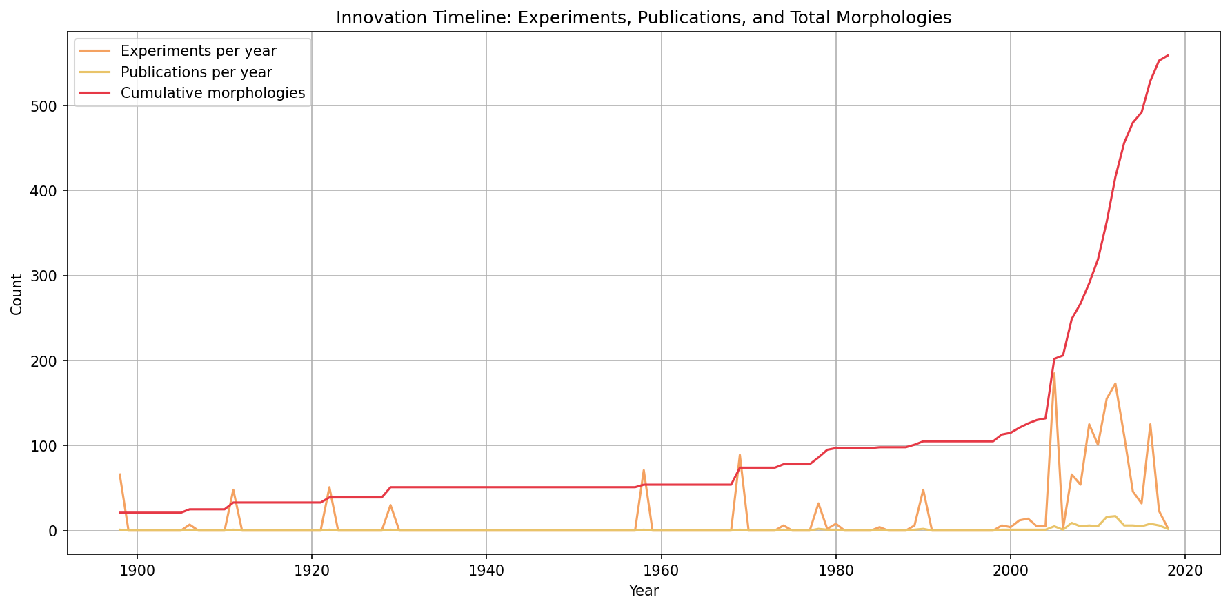
4 · The corpus the grammar was trained onPlanformDB · Lobo / Malone / Levin · ~16 decades of cut-and-graft
Real output of 1900-planformDB_parser.py from fractastical/morpholib. The 2015 evolutionary search ran against this corpus — every documented experiment, every observed morphology, machine-readable.
19 / 33
Slide 20 · Same cells, different body
2020–21 · Kriegman, Blackiston, Bongard, Levin et al.
If form is partly software, you can recompile it — the xenobot.
Take embryonic skin cells from Xenopus laevis, remove the rest of the embryo, let the cells
reaggregate. They do not become frog skin.
They self-organise into a novel anatomy:
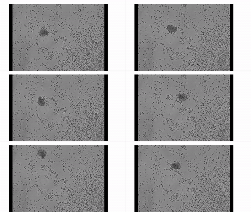
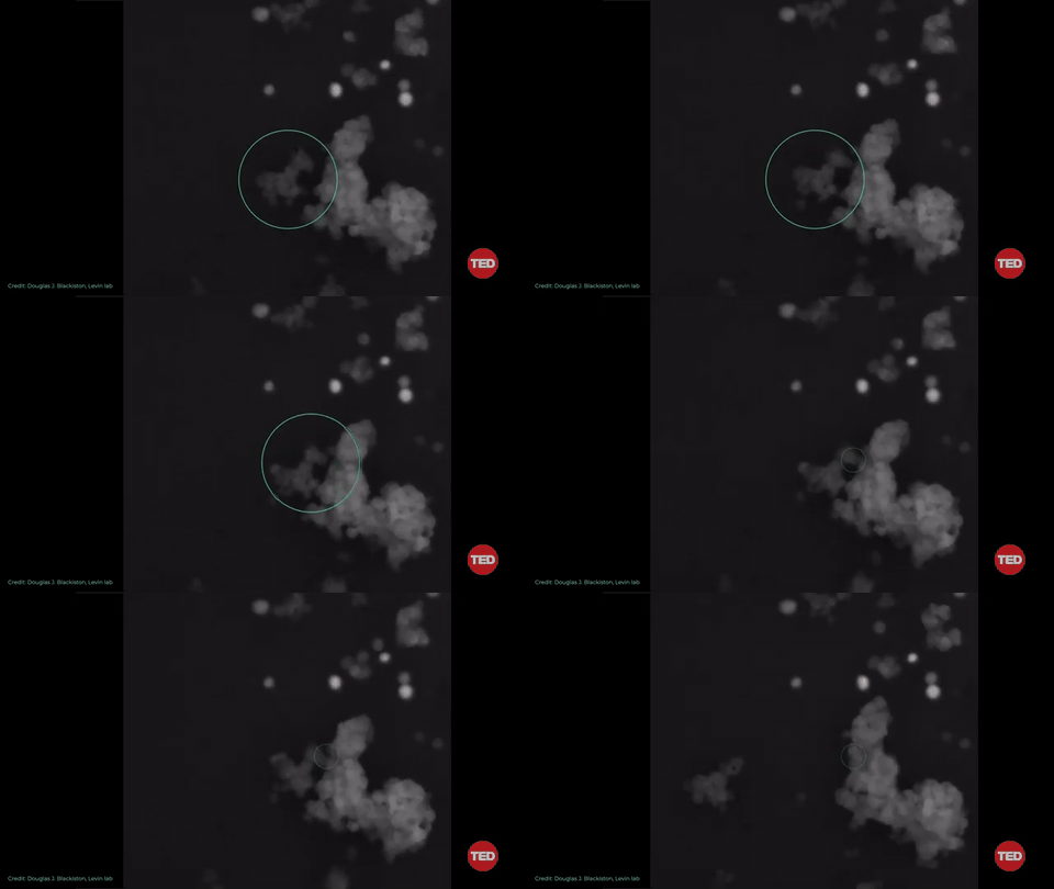
2020 — first xenobots, designed by an evolutionary algorithm and assembled by hand
(Kriegman et al., PNAS).
2021 — ciliated, self-propelling forms (Blackiston et al., Sci. Robotics) and
kinematic self-replication, where xenobots gather loose cells into new xenobots
(Kriegman et al., PNAS).
The genome doesn't encode a frog. It encodes capacity.
Xenobot navigating a field of loose cells. Source: M. Levin / Allen Discovery Center, Tufts.
20 / 33
Slide 21 · Built from human cells
2024 · Gumuskaya et al., Advanced Science
Anthrobots interact with human tissue in culture.
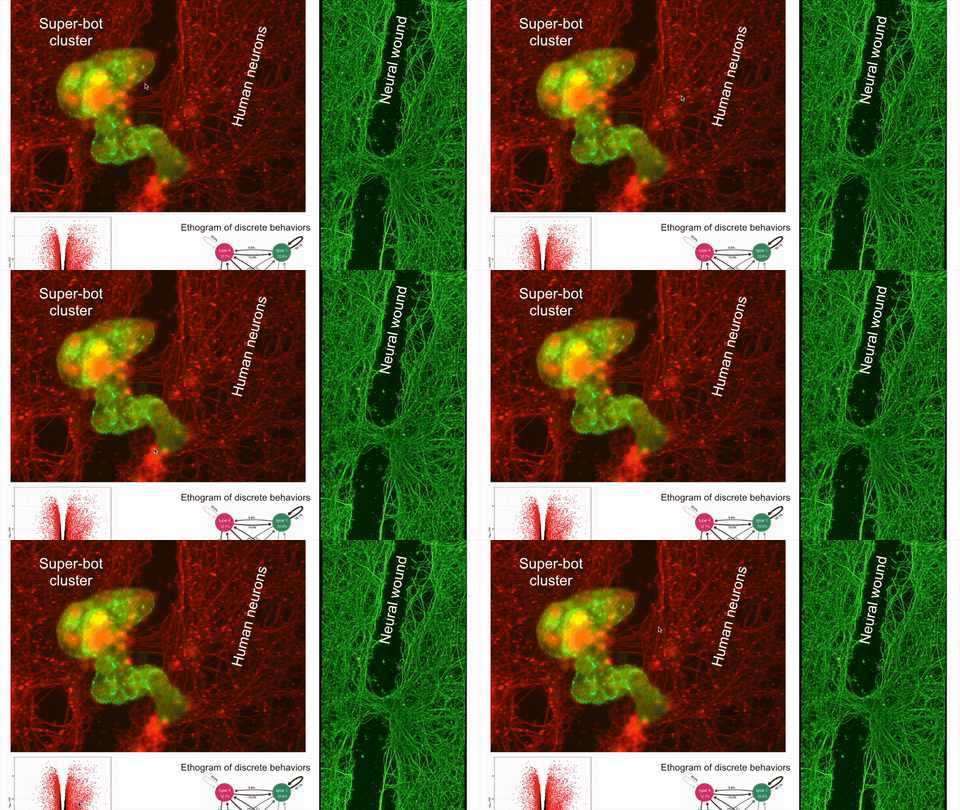
Made from adult human tracheal cells, anthrobots aggregate into "super-bot" clusters. In
in vitro assays they move across cultured human neurons, congregate near injuries, and
are associated with improved closure in neuronal-wound assays.
Because they're built from the same cells as the tissue around them, they natively share its
bioelectric and biochemical signalling — making them candidate research tools
for studying repair and tissue dynamics.
Super-bot cluster (green) on cultured human neurons (red), next to a neural wound. Source: Gumuskaya et al. (2024), Levin lab.
21 / 33
Slide 22 · The anatomical compiler
Murugan et al. (Levin lab) 2022 · Science Advances · 18-month follow-up in adult Xenopus laevis
Specify the anatomy. Let the cells compile it.
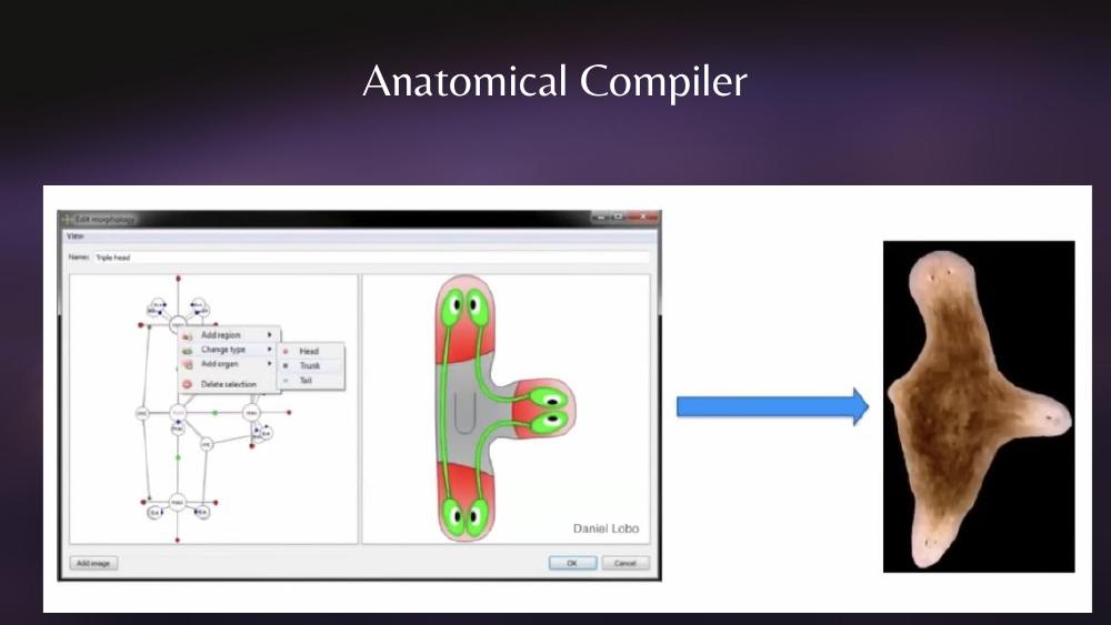
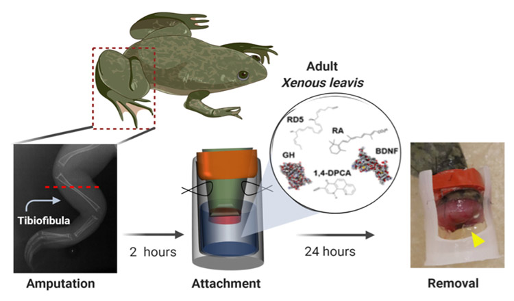
Stop micromanaging tissues; hand cells a target morphology and let them execute. Lobo & Levin built a literal compiler — a graphical model where you draw the body plan you want and the planarian regenerates it. A 24-hour drug cocktail in a wearable silicone BioDome then carries the same idea into adult Xenopus: 18 months of patterned limb outgrowth from one intervention.
● Compiler · Lobo & Levin · planarian regeneration model
Specify the target body plan in the editor (here: triple-head). The planarian regenerates that shape. The compiler turned "anatomical compiler" from metaphor into a literal piece of software.
● Compiler in vivo · Murugan et al. 2022 · BioDome on adult Xenopus
Adult X. laevis hindlimb amputation → silicone BioDome cap loaded with a 5-drug cocktail (BDNF · 1,4-DPCA · RD5 · GH · RA) → 24 h drug delivery → cap off. Bone, vasculature, innervation and motor function all show up over the next 18 months. Murugan et al., Sci. Adv. 2022.
The same excitable-cell layer that builds bodies can also be wired up to compute.
Bioelectricity becomes the I/O channel — voltage in, voltage out — and the cells
do the work in between.
DishBrain (Kagan et al., Neuron 2022) — ~800 K mouse + human-iPSC (induced pluripotent stem cells: adult cells reprogrammed back to a stem-cell state) cortical neurons on a high-density MEA(multi-electrode array — a chip with hundreds of micro-electrodes the cells grow on, used for both reading and stimulating activity), taught to play Pong via closed-loop electrical feedback — the authors interpret the training scheme through the Free-Energy Principle(Friston's framework: systems learn by minimizing prediction error / "surprise"), though the implementation itself is heuristic closed-loop feedback.
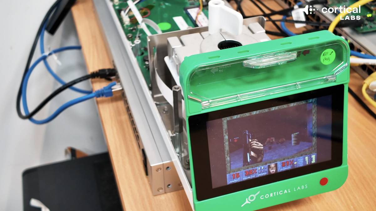
Cortical Labs · CL1 (Melbourne, launched 2025) — billed as the first commercially available biological computer; cloud-accessible "Synthetic Biological Intelligence" platform building on the lab's earlier DishBrain hardware (partner access from 2023–24).
FinalSpark Neuroplatform (Vevey, Switzerland; Neuroplatform publicly described 2023) — remote API access to ~16 living brain organoids(mm-scale 3D self-organized neural tissue grown from stem cells — "mini-brains"); Wetware-as-a-Service.
Brainoware (Cai et al., Nature Electronics 2023) — brain organoid used as the reservoir in reservoir computing(a network whose fixed, rich dynamics do most of the work; only a small readout layer is trained); speech-recognition demo.
Why this slide is here: every prior slide treated bioelectricity as a morphogenetic
signal. These groups treat the same circuitry as a computational one — a parallel
frontier with shared physics, shared tools (MEAs, GEVIs, optogenetic ion channels), and
a very different end-game.
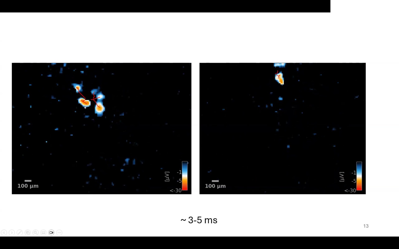
Closed-loop MEA + cultured cortical neurons · the substrate underneath every group on this slide.
Cortical Labs CL1 (2025) running DOOM · 200 K living human neurons driving the gameplay. Credit: Cortical Labs.
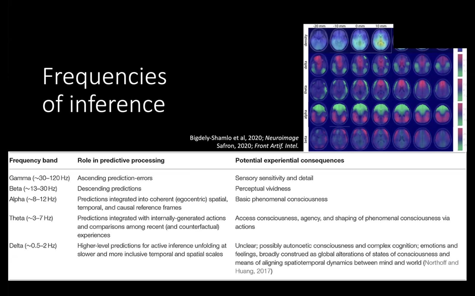
Different EEG bands = different scales of prediction.
If cognition is hierarchical inference, each oscillatory band carries a different layer of the predictive stack: gamma is the ascending error signal, beta the descending prediction, alpha integrates them into a frame, theta brings agency in, delta sets the slow context. Bioelectricity isn't just resting potentials — it's computational potentials at multiple harmonics simultaneously.
24 / 33
Slide 25 · The cell palette
Survey · model systems used in bioelectricity & synthetic morphology
The same logic, across very different cells.
A small zoo of model systems — each stress-tests the same claim (tissue-level voltage instructs anatomy) in a different cell type and body plan.
Two-headed regeneration via gap-junction perturbation (2010–13); pattern memory persists through fission (Durant 2017).
Human airway
Tracheal epithelial cells
Anthrobots — adult human cells self-organize into motile multicellular bots and heal neuronal wounds in vitro (Gumuskaya 2024).
Human cortex
iPSC-derived neurons
The substrate anthrobots interact with — provides the readout for behavioural / wound-healing assays in the 2024 work.
Axolotl & tadpole
Regeneration models
Limb-regrowth with bioelectric drug cocktails on Xenopus tadpoles (Murugan / Levin 2022); axolotl as a comparative reference.
Cell lines & cancer
Melanoma, glia, MDCK
Bioelectric perturbation triggers a neoplastic-like conversion of melanocytes in Xenopus embryos (Blackiston et al. 2011); cancer as a tissue-voltage failure mode.
25 / 33
Slide 26 · From scientist-in-the-loop to robot scientist
Tool · MomBot · Levin lab, Tufts
An AI brain + a robot body, running cell experiments around the clock.
Closed-loop autonomous labs have already proven themselves in chemistry (LBNL's A-Lab) and
pharma (Strateos, Emerald Cloud). The Levin lab brought the same idea to living cells.
MomBot — the Tufts prototype. First proof a bioelectric experiment could be designed, run and scored on a closed loop with no human in the seat.
One of the few AI + wet-lab systems explicitly aimed at living-cell collective behaviour, not just chemistry or pharma screens.
Pairs an AI experiment-design "brain" with robotic plate-handling, imaging and stimulation — every result feeds the next hypothesis.
Closed-loop wet-lab automation · the substrate MomBot pioneered for living-cell experiments.
26 / 33
Slide 27 · The labs landscape
Survey · 9 featured labs + ~75 tracked PIs
The field is distributed.
Pattern formation · regeneration
Levin Lab · Allen Discovery Center
Tufts University
Bioelectric control of anatomy — two-headed planaria, xenobots, anthrobots, BioDome.
Bio-inspired engineering
Wyss Institute
Harvard University
Pattern memory, AI top-down regulation, multiplexed Xenopus culture, therapeutics.
Computer-designed organisms
ICDO
Tufts & University of Vermont
Levin × Bongard joint institute — life-machine hybrids, regen medicine, cancer targeting.
Computational morphogenesis
Lobo Lab
UMBC
Automated discovery of regulatory networks for planarian regeneration.
Evolutionary robotics
Bongard Lab · Proteus
University of Vermont
Xenobot co-creator — morphological computation, co-evolved brains and bodies.
Mechanobiology · organs-on-chips
Ingber Lab
Wyss Institute · Harvard
Organs-on-chips, tissue mechanics, the engineering arm at Wyss.
Voltage imaging tools
Cohen Lab
Harvard University
Optopatch, Archon, the GEVI toolkit that lets you film a cell think.
Bioelectric oncology
Gentile Lab
Medical Univ. of South Carolina
K+-channel activators (NS1643) → senescence in breast and colon cancer.
Active inference · basal cognition
Pezzulo Group
CNR · Rome
Anticipatory cognition — predictive frame for cells, tissues and bodies.
…and the wider network around them — ~75 PIs the paper scanner watches each week (watchlist.ts)
Levin Lab core · 39 active
Michael Levin, Patrick McMillen, Vaibhav Pai, Wesley Clawson, Hananel Hazan, Franz Kuchling, Juanita Mathews, Federico Pigozzi, Yanbo Zhang, Caitlin Grasso, Jack Lindsay, Jayati Mandal, Patrick Erickson, Tomáš Pelikán, Doug Blackiston · + 24 more
Voltage imaging & tooling
Adam Cohen (Harvard · GEVIs), Ed Boyden (MIT · Archon), Eve Marder (Brandeis), Donald Ingber (Wyss)
Wound currents & channelopathies
Min Zhao (UC Davis), Mustafa Djamgoz (Imperial), Richard Nuccitelli, Christine Pullar (Leicester), Colin McCaig, Ann Rajnicek (Aberdeen), Richard H. W. Funk (TU Dresden)
Saverio Gentile (MUSC · Kv11.1 / NS1643), Mustafa Djamgoz (voltage-gated Na in metastasis)
Morphogenesis · dev-bio
Stuart A. Newman (NYMC), Aneta Koseska (MPI), Matthew Harris (Harvard), Koji Tamura (Tohoku), Bassem Hassan (ICM Paris), Kelly McLaughlin (Tufts)
Basal cognition · unconventional substrates
František Baluška (Bonn), Stefano Mancuso (Florence), Audrey Dussutour (CNRS · slime moulds), Eshel Ben-Jacob, Andy Adamatzky (UWE · fungal computing)
Theory · active inference · xenobot collaborators
Karl Friston (UCL), Chris Fields, Josh Bongard (UVM · xenobot co-author), Sebastian Risi (ITU · neural CA), Giovanni Pezzulo (CNR)
Cross-disciplinary collaborators
Daniel Ari Friedman (Active Inference Institute), Doug Blackiston (Tufts · xenobot construction lead) — plus Wyss / ICDO / Allen-Discovery-Center engineering staff.
27 / 33
Slide 28 · The latent platonic space
Concept · Levin & Hazan symposium 2025
Raup's morphospace, generalized.
If voltage maps can navigate cells through the space of possible bodies, where does that space
live? Levin's working answer: a structured, non-physical space of forms, patterns and
higher agencies that biological systems (and minds, and AI) appear to discover, not invent.
Attractor landscape
Anatomies are stable basins the system gravitates toward. Bioelectric circuits encode which.
Active inference
Cells minimize surprise against a target morphology — Friston's framework, applied to shape.
Cognitive platonism
Forms are real as operational constraints, not as transcendent objects (Dodig-Crnkovic).
Convergence
Biology, cognition and large-scale ML all seem to land on overlapping patches of this space.
Vmem wave propagating across a tissue grid · inspired by BETSE bioelectric simulations (Pietak & Levin 2016).
28 / 33
Slide 29 · A use case: bioelectric oncology
2013–present · Saverio Gentile · Medical University of South Carolina
Open a K+ channel. Stop a tumour.
Everything so far has been biology. Saverio Gentile's lab at MUSC is the clearest existing demonstration that the bioelectric layer is also directly druggable in the clinic — specifically, in cancer.
The drug class: small-molecule activators of the hERG / Eag (KCNH) K+ channel family — most prominently NS1643. Opening these channels lets K+ efflux, hyperpolarizes the cancer cell, and pushes it out of the cell cycle.
The result: Lansu & Gentile, Cell Death & Disease (2013) — NS1643 induces p21-mediated cellular senescence in MDA-MB-231 triple-negative breast cancer cells. The cells stop dividing, permanently, with no classical cytotoxic killing. The same program follows in HCT-116 colorectal cancer.
In vivo: Gentile-lab follow-up work shows mouse-xenograft tumour-mass reduction and synergy with standard chemotherapy.
Why this is the clinical translation: NS1643 and its cousins are already-characterized molecules originally developed against cardiac arrhythmia. The "electroceutical" framing (Tuszynski, Tilli & Levin 2017) becomes a concrete, repurposable drug class — bioelectricity moves from basic science to oncology pipeline.
The connecting thread: upstream — Blackiston & Levin 2011 (depolarization drives metastasis with no oncogene) and Chernet & Levin 2013/2016 (forced hyperpolarization suppresses KRAS tumours in Xenopus embryos). Downstream — Gentile turns that biology into a drug.
A depolarized cancer-like cluster (red) hyperpolarized back to healthy (teal) by re-opening K+ channels — the Gentile lab's pharmacological premise.
29 / 33
Slide 30 · The live map
Tool · ProbKnow · live literature graph
The bioelectricity literature, graphed in real time.
ProbKnow ingests new papers, links them to ~2,700 testable predictions, and lights up the hypothesis lanes as evidence arrives. Live tool → probknow.com.
30 / 33
Slide 31 · The through-line
One idea, eight steps, a hundred years.
Each step makes the same claim sharper: genes are the parts list, but something at the tissue
scale helps decide what gets built — and bioelectricity is a major part of that something.
What each step added
1917 — Thompson: there is a field, mathematically
1952 — Turing: the math alone predicts patterns from a uniform field
1966 — Raup: form lives in a space, mostly empty
2011 — Vandenberg, Morrie & Adams: the field is visible as voltage
2010–17 — Levin lab: the field is editable, and edits can persist
2013–present — Gentile lab: a K+ channel activator (NS1643) induces senescence in breast and colon cancer — bioelectricity becomes a drug
2020–21 — Kriegman et al.: cells can be coaxed to build novel bodies
2024 — Gumuskaya et al.: those bodies can interact with human tissue in culture
Why it matters for the conference
Read/write tools for anatomy — voltage dyes, channel openers/blockers
Regenerative medicine — pattern memory as a therapeutic target
Developmental biology — a tissue-scale layer above gene regulation
The genome encodes the hardware. Bioelectricity is part of how cells decide what to do with it.
31 / 33
Slide 32 · Sources & further reading
Where each step comes from
1780–1952 — Discovery of bioelectricity
Galvani, De Viribus Electricitatis in Motu Musculari Commentarius (1791). Volta, Phil. Trans. R. Soc. (1800) on the voltaic pile. Du Bois-Reymond, Untersuchungen über thierische Elektricität (1848–84). Bernstein, "Untersuchungen zur Thermodynamik der bioelektrischen Ströme," Pflügers Archiv (1902). Hodgkin & Huxley, "A quantitative description of membrane current and its application to conduction and excitation in nerve," J. Physiol. 117 (1952). Galvani plate: public domain via De Viribus Electricitatis Tab. I.
1917 — D'Arcy Thompson On Growth and Form, Cambridge University Press. Animation by S. M. Blinder, Wolfram Demonstrations Project.
1952 — Alan Turing
Turing AM, "The Chemical Basis of Morphogenesis," Philosophical Transactions of the Royal Society B 237 (1952). Confirmations: Kondo S & Asai R, "A reaction-diffusion wave on the skin of the marine angelfish Pomacanthus," Nature 376 (1995); Sick S et al., "WNT and DKK determine hair follicle spacing through a reaction-diffusion mechanism," Science 314 (2006); Sheth R et al., "Hox genes regulate digit patterning by controlling the wavelength of a Turing-type mechanism," Science 338 (2012). Bioelectric extension: Pietak A & Levin M, "Exploring instructive physiological signaling with the Bioelectric Tissue Simulation Engine (BETSE)," Frontiers in Bioengineering & Biotechnology 4: 55 (2016).
1966 — David Raup
"Geometric Analysis of Shell Coiling," J. Paleontology. Animation by Phil Ramsden, Wolfram Demonstrations Project.
Mechanism primer
Levin, "Bioelectric signaling: Reprogrammable circuits underlying embryogenesis, regeneration, and cancer," Cell (2021). Levin & Martyniuk, "The bioelectric code," BioSystems (2018).
2011 — Face of a Frog
Vandenberg LN, Morrie RD, Adams DS, "V-ATPase-dependent ectodermal voltage and pH regionalization are required for craniofacial morphogenesis," Developmental Dynamics 240 (2011). Earlier mechanistic work: Adams DS et al., Development (2006).
2010–17 — Two-headed planaria
Oviedo NJ et al., Dev. Biol. (2010); Beane WS et al., Chem. & Biol. (2011); Durant F et al., Biophys. J. (2017) — long-term maintenance of the altered head/tail target morphology across re-amputation, via stable gap-junctional state changes.
2013–present — Use case: bioelectric oncology (Gentile lab) Lansu K & Gentile S, "Potassium channel activation inhibits proliferation of breast cancer cells by activating a senescence program," Cell Death & Disease 4:e652 (2013) — NS1643 / hERG activator induces p21-mediated senescence in MDA-MB-231 triple-negative breast cancer; subsequent Gentile-lab work extends the program to HCT-116 colorectal cancer and to mouse xenograft tumour-mass reduction. Upstream basis: Blackiston DJ, Adams DS, Lemire JM, Lobikin M, Levin M, "Transmembrane potential of GlyCl-expressing instructor cells induces a neoplastic-like conversion of melanocytes via a serotonergic pathway," Disease Models & Mechanisms 4 (2011); Blackiston DJ & Levin M, "Bioelectric controls of cell proliferation: ion channels, membrane voltage and the cell cycle," Cell Cycle 10 (2011); Chernet BT & Levin M, "Endogenous voltage potentials and the microenvironment," J. Clinical & Experimental Oncology S1 (2013); Chernet BT, Adams DS, Pai VP, Levin M, "Use of genetically encoded, light-gated ion translocators to control tumorigenesis," Oncotarget 7 (2016) — KRAS-driven tumour suppression via forced hyperpolarization. Framing: Tuszynski J, Tilli TM & Levin M, "Ion channel and neurotransmitter modulators as electroceutical approaches to the control of cancer," Current Pharmaceutical Design 23 (2017). Adjacent drug-repurposing line: Brackenbury WJ & Djamgoz MBA on Nav-channel-driven cancer invasion (J. Physiol. 573, 2006); Djamgoz MBA & Onkal R on Nav blockers as anti-metastatic agents (Recent Patents on Anti-Cancer Drug Discovery 8, 2013).
2020–21 — Xenobots
Kriegman S et al., "A scalable pipeline for designing reconfigurable organisms," PNAS 117 (2020); Blackiston D et al., "A cellular platform for the development of synthetic living machines," Sci. Robotics 6 (2021); Kriegman S et al., "Kinematic self-replication in reconfigurable organisms," PNAS 118 (2021).
2024 — Anthrobots
Gumuskaya G et al., "Motile living biobots self-construct from adult human somatic progenitor seed cells," Advanced Science 11 (2024).
Cell palette — additional sources
Blackiston DJ, Adams DS, Lemire JM, Lobikin M, Levin M, "Transmembrane potential of GlyCl-expressing instructor cells induces a neoplastic-like conversion of melanocytes via a serotonergic pathway," Disease Models & Mechanisms 4 (2011); Blackiston DJ & Levin M, "Bioelectric controls of cell proliferation: ion channels, membrane voltage and the cell cycle," Cell Cycle 10 (2011).
Murugan NJ et al. (Levin lab), "Acute multidrug delivery via a wearable bioreactor facilitates long-term limb regeneration and functional recovery in adult Xenopus laevis," Science Advances 8 (2022) — bioelectric / drug-cocktail tadpole limb regrowth.
Latent platonic space
Levin M & Hazan H, symposium & working notes (2025) on the latent platonic space of forms and agencies; see Levin, "Self-improvising memory: a perspective on memories as agential, dynamically reinterpreting cognitive glue," Entropy 26 (2024) for the surrounding cognitive-platonism framing. Glossary: /platonic-space.
Biocomputers — DishBrain · CL1 · FinalSpark · Brainoware
Kagan BJ, Kitchen AC, Tran NT, et al., "In vitro neurons learn and exhibit sentience when embodied in a simulated game-world," Neuron 110, 3952–3969 (2022) — the original DishBrain Pong result. Cortical Labs CL1 (Melbourne, launched 2025) — billed as the first commercially available biological computer; builds on the lab's earlier DishBrain hardware (partner access from 2023-24); see corticallabs.com. FinalSpark Neuroplatform — remote-access bio-computing on living brain organoids; Vevey, Switzerland; Neuroplatform publicly described 2023; see finalspark.com. Cai H, Ao Z, Tian C, et al., "Brain organoid reservoir computing for artificial intelligence," Nature Electronics 6, 1032–1039 (2023) — Brainoware speech-recognition demo.
Bioelectric Code of Life — live workshop
SEMF Workshop Series, 7 March 2026 (online). Frontier seminar bridging bioelectric theory with applications. Registration & programme: bioelectricitycourse.com.
Original animations & reconstructions
Slide 06 (frog-leg twitch): original inline SVG depicting Galvani's bimetal-arc circuit closure (1791). Slide 10 (cell sorting): inline SVG inspired by Steinberg's differential adhesion hypothesis, Science (1963), and the Glazier-Graner cellular Potts model, Phys. Rev. Lett. (1992); Morpheus M0021 reference implementation. Slide 11 (mechanism): original inline SVG of ions / gap-junction equilibration / Vmem → anatomy. Slide 13 (imaging-toolkit voltage trace) and Slide 14 (timeline scrubber): original CSS/SVG. Slide 22 (anatomical compiler): original inline SVG of the BioDome regeneration timeline (amputation → BioDome → blastema → patterned limb). Slide 25 (cell-palette montage): stitched from CC-licensed lab clips referenced above. Slide 29 (Vmem wave on tissue grid): inline SVG inspired by BETSE — Pietak A & Levin M, "Exploring instructive physiological signaling with the Bioelectric Tissue Simulation Engine," Frontiers in Bioengineering and Biotechnology 4: 55 (2016), CC BY; source code BSD-2 at gitlab.com/betse/betse.
Bioelectricity Nexus bioelectricitynexus.com — curated hub for papers, researchers, and tools.
32 / 33
Slide 33 · The last word
Michael Levin · SEMF 2026 · closing remarks
Over to Mike Levin.
Audio plays through the autoplay bar ↓
33 / 33
Appendix A1 · Why this took so long
D'Arcy Wentworth Thompson · On Growth and Form · 1917 · public domain
A century-old answer to "why isn't morphology already mathematical?"
Thompson saw, before any of the modern tools existed, exactly the obstacle this deck has been pushing against — and pointed at the way out. Three sentences from page 642:
"The details in which the figure differs from its mathematical prototype are more important and more interesting than the features in which it agrees."
"No chain hangs in a perfect catenary and no raindrop is a perfect sphere; and this for the simple reason that forces and resistances other than the main one are inevitably at work."
"The same is true of organic form, but it is for the mathematician to unravel the conflicting forces which are at work together."
A1 · appendix
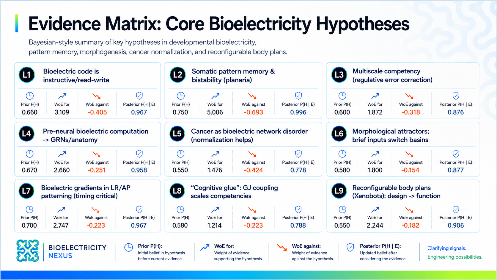
Appendix A2 · Evidence matrix
L1 → L9 · Bayesian-style summary
Nine core hypotheses · prior, evidence weight, posterior
A2 · appendix
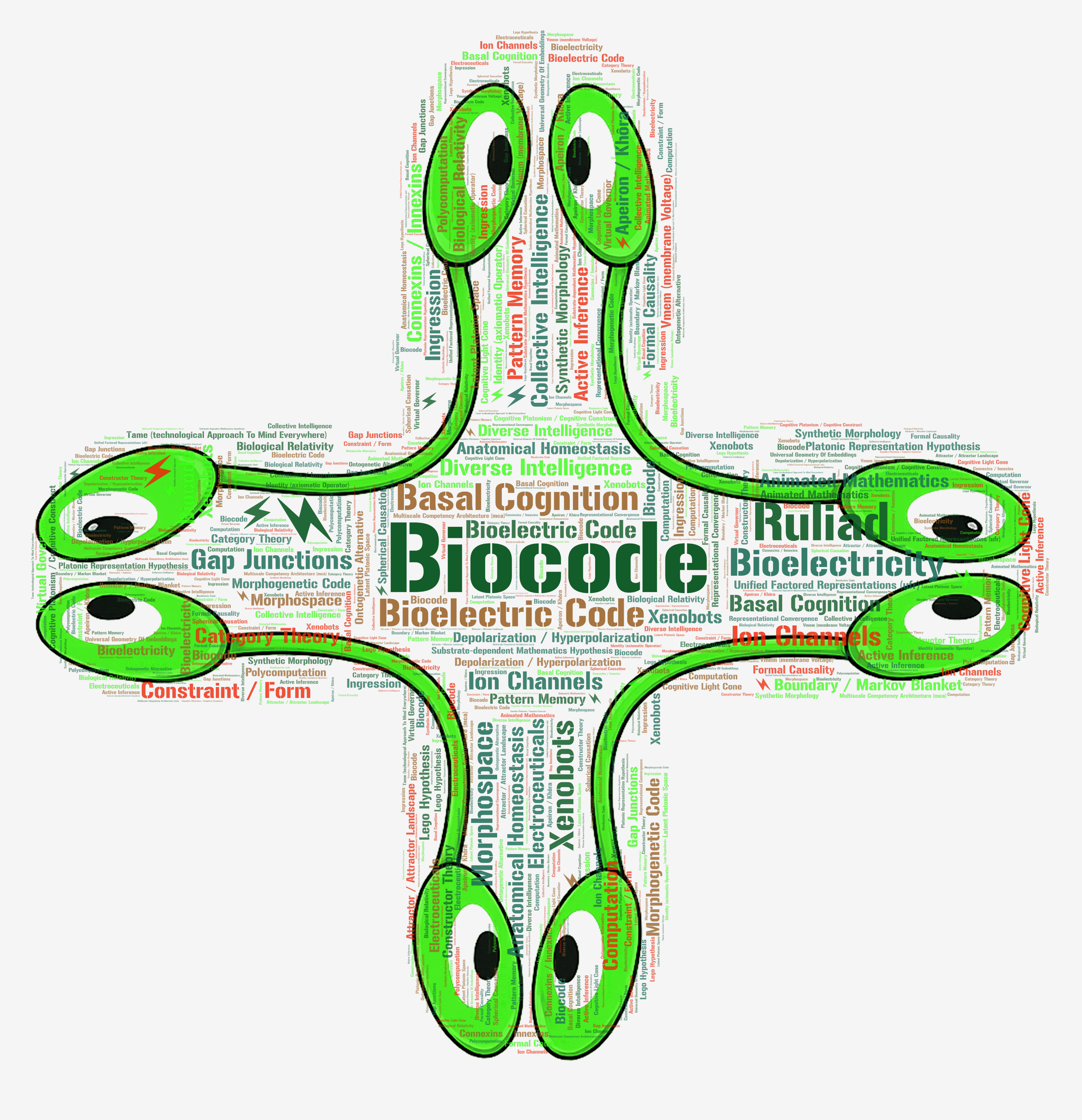
Appendix A3 · Biocode word-cloud
Click any term · jumps to its glossary card on slide 5
The whole vocabulary, in one cell-shaped sticker.
Click any term · opens the full /glossary at that entry